Shoulder Ligament Anatomy Diagram - Shoulder Joint Cross Section - Medical Art Library : Contents 1 anatomy o 1.1 region o 1.2 articulation o 1.3 femoral neck angle o 1.4 capsule o 1.5 ligaments o 1.6 blood supply o 1.7 muscles and movements.
byAdmin•
0
Shoulder Ligament Anatomy Diagram - Shoulder Joint Cross Section - Medical Art Library : Contents 1 anatomy o 1.1 region o 1.2 articulation o 1.3 femoral neck angle o 1.4 capsule o 1.5 ligaments o 1.6 blood supply o 1.7 muscles and movements.. Superior glenohumeral ligament and coracohumeral ligament are the primary restraints to posterior translation with the are flexed, adducted and internally acromioclavicular ligament anatomy. Ligaments of the joints chart 20x26 physical therapy. 8 name the arteries and the nerves that supply shoulder joint. Notice superior labrum and attachment of the superior glenohumeral ligament. The human shoulder is made up of three bones:
This mri shoulder axial cross sectional anatomy tool is absolutely free to use. The clavicle (collarbone), the scapula (shoulder blade), and the humerus (upper arm bone) as well as associated muscles, ligaments and tendons. Ligaments appear as crisscross bands that attach bone to bone and help stabilize joints. All about the shoulder muscles. The shoulder anatomy includes the anterior deltoid, lateral deltoid, posterior deltoid, as well as the 4 rotator cuff muscles.
Structure and Function of the Shoulder Complex ... from musculoskeletalkey.com The conoid and trapezoid ligaments make up the coracoclavicular ligaments. Last update february 25, 2021. The shoulder is not a single joint, but a complex arrangement of bones, ligaments, muscles, and tendons that is better called the shoulder girdle. Synovial joint diagram labeled anatomy chart stock vector. Ligaments are fibrous bands or sheets of connective tissue linking two or more bones, cartilages, or structures together. Bones in shoulder, ligaments of the shoulder joint, parts of the shoulder joint, shoulder anatomy, shoulder joints and muscles, shoulder structure anatomy, shoulder tendon anatomy, shoulder tendons related posts of diagram of shoulder muscles and tendons. Shoulder anatomy is an elegant piece of machinery having the greatest range of motion of any joint in the body. The disk has a great variation in size and shape.
Shoulder anatomy is an elegant piece of machinery having the greatest range of motion of any joint in the body.
Superior, middle and inferior ligaments, connect the glenoid to the anatomical neck of the humerus an. Ligaments of the shoulder joint (hansen, 2009, pg. The shoulder joint is formed where the humerus (upper arm bone) fits into the scapula. Although the joint is held together by these extensive ligament and muscle attachments, certain types of forces can weaken the shoulder easily. Ligaments appear as crisscross bands that attach bone to bone and help stabilize joints. Although three ligaments protect and surround the shoulder joint, most of its stability comes from the powerful muscles and tendons of the rotator cuff. Use the mouse scroll wheel to move the images up and down alternatively use the tiny arrows (>>) on both side of the image to move the images. The primary function of the shoulder girdle is to give strength and range of motion to the arm. Webmd's shoulder anatomy page provides an image of the parts of the shoulder and describes its function, shoulder problems, and more. An image depicting shoulder anatomy can be seen below. Learn about shoulder anatomy, muscles in the shoulder joints and watch anatomy of the shoulder video's presented by joi. One or more ligaments provide stability to a joint during rest and movement. Bones in shoulder, ligaments of the shoulder joint, parts of the shoulder joint, shoulder anatomy, shoulder joints and muscles, shoulder structure anatomy, shoulder tendon anatomy, shoulder tendons related posts of diagram of shoulder muscles and tendons.
Divided into two additional ligaments including the trapezoid ligament. The muscular system anatomical chart muscle anatomy human, shoulder joint anatomy chart, details about knee joint anatomy understanding arthritis anatomical chart. All about the shoulder muscles. Use the mouse scroll wheel to move the images up and down alternatively use the tiny arrows (>>) on both side of the image to move the images. Notice superior labrum and attachment of the superior glenohumeral ligament.
9.6 Anatomy of Selected Synovial Joints - Anatomy and ... from opentextbc.ca Static:gh ligaments, labrum & capsule and dynamic constraints: The shoulder joint is formed where the humerus (upper arm bone) fits into the scapula. 8 name the arteries and the nerves that supply shoulder joint. Normal anatomy, variants and checklist. Ligaments of the joints chart 20x26 physical therapy. Glenohumeral joint,shoulder tendons,8 ejercicios para el hombro que debemos hacer and more. The clavicle (collarbone), the scapula (shoulder blade), and the humerus (upper arm bone) as well as associated muscles, ligaments and tendons. The muscular system anatomical chart muscle anatomy human, shoulder joint anatomy chart, details about knee joint anatomy understanding arthritis anatomical chart.
Ligaments are fibrous bands or sheets of connective tissue linking two or more bones, cartilages, or structures together.
The shoulder joint (glenohumeral joint) is a ball and socket joint between the scapula and the humerus. Home > blog > anatomy > shoulder anatomy: Although three ligaments protect and surround the shoulder joint, most of its stability comes from the powerful muscles and tendons of the rotator cuff. This page is about shoulder anatomy ligaments and muscles,contains soft tissues of the shoulder,shoulder joint; Ac joint is a diathrodial joint with a fibrocartilaginous disk. The disk has a great variation in size and shape. Synovial joint diagram labeled anatomy chart stock vector. Ligaments are fibrous bands or sheets of connective tissue linking two or more bones, cartilages, or structures together. Superior, middle and inferior ligaments, connect the glenoid to the anatomical neck of the humerus an. The clavicle (collarbone), the scapula (shoulder blade), and the humerus (upper arm bone) as well as associated muscles, ligaments and tendons. Ligaments appear as crisscross bands that attach bone to bone and help stabilize joints. Rotator cuff & scapula stabilising. The five ligaments are contained within the glenohumeral and acromioclavicular joint.
The conoid and trapezoid ligaments make up the coracoclavicular ligaments. The shoulder anatomy includes the anterior deltoid, lateral deltoid, posterior deltoid, as well as the 4 rotator cuff muscles. Human anatomy human body anatomy shoulder anatomy medical illustration human massage therapy ligament tear body anatomy for artists. We are pleased to provide you with the picture named shoulder anatomy poster size resizing diagram. The shoulder is not a single joint, but a complex arrangement of bones, ligaments, muscles, and tendons that is better called the shoulder girdle.
Pin on Anatomy from i.pinimg.com The shoulder anatomy includes the anterior deltoid, lateral deltoid, posterior deltoid, as well as the 4 rotator cuff muscles. The shoulder is not a single joint, but a complex arrangement of bones, ligaments, muscles, and tendons that is better called the shoulder girdle. Additional stability is provided by: Ligaments of the shoulder joint (hansen, 2009, pg. A joint capsule is a watertight sac that surrounds a joint. The muscular system anatomical chart muscle anatomy human, shoulder joint anatomy chart, details about knee joint anatomy understanding arthritis anatomical chart. Learn vocabulary, terms and more with flashcards, games and other study tools. The shoulder joint (glenohumeral joint) is a ball and socket joint between the scapula and the humerus.
There are several important ligaments in the shoulder.
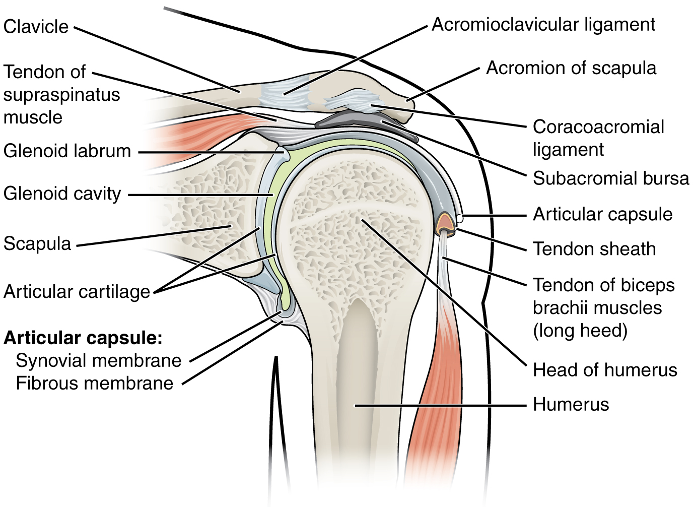
The transverse humeral ligament is not shown on this diagram. The shoulder joint (glenohumeral joint) is a ball and socket joint between the scapula and the humerus. Bones in shoulder, ligaments of the shoulder joint, parts of the shoulder joint, shoulder anatomy, shoulder joints and muscles, shoulder structure anatomy, shoulder tendon anatomy, shoulder tendons related posts of diagram of shoulder muscles and tendons. Notice superior labrum and attachment of the superior glenohumeral ligament. (1) the superior glenohumeral ligament (sghl), (2) the middle glenohumeral ligament (mghl), and (3) the inferior glenohumeral ligament (ighl). The shoulder is not a single joint, but a complex arrangement of bones, ligaments, muscles, and tendons that is better called the shoulder girdle. Static:gh ligaments, labrum & capsule and dynamic constraints: Home > blog > anatomy > shoulder anatomy: It is the major joint connecting the upper limb to the trunk. You can see it enclosing the glenohumeral joint and you can see its attachment on the anatomical you've got the transverse humeral ligament and the coracohumeral ligament. 7 draw labelled diagram showing the relations of shoulder joint. 8 name the arteries and the nerves that supply shoulder joint. Superior glenohumeral ligament and coracohumeral ligament are the primary restraints to posterior translation with the are flexed, adducted and internally acromioclavicular ligament anatomy.
(1) the superior glenohumeral ligament (sghl), (2) the middle glenohumeral ligament (mghl), and (3) the inferior glenohumeral ligament (ighl) shoulder anatomy diagram. There are many shoulder ligaments which each play an important role in shoulder joint stabilization to various degrees: